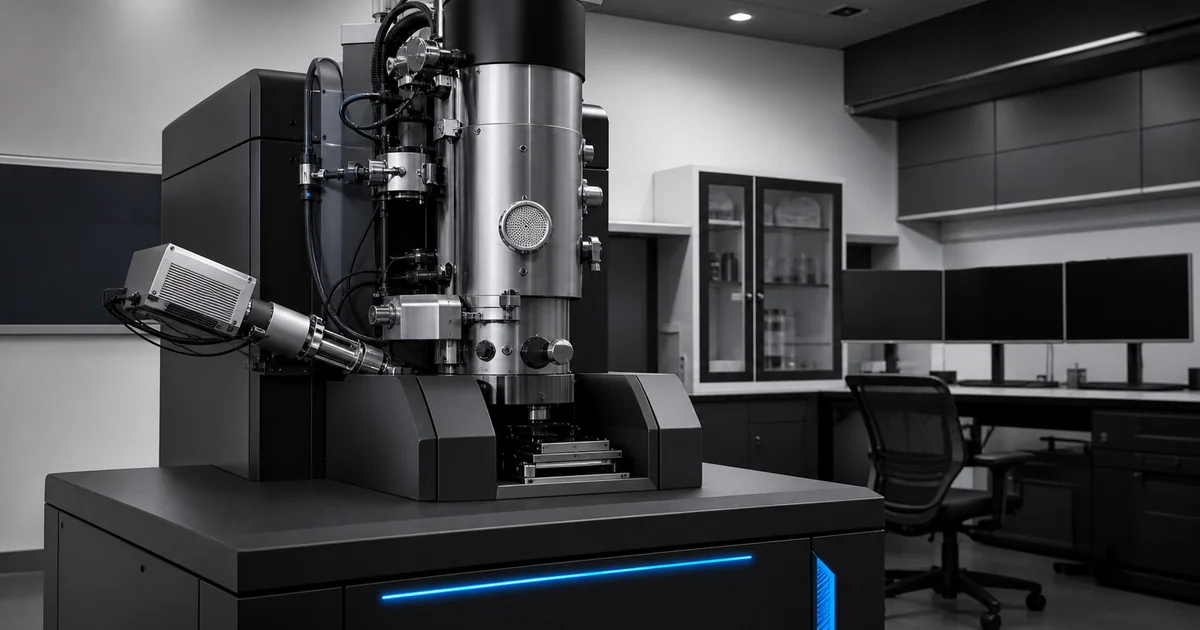
Quick answer
SEM resolution is the ability to separate fine details. SEM magnification is how large the scanned area appears on the display. Vacuum is the low pressure environment that lets electrons travel through the instrument and interact with the sample in a controlled way.
Good SEM imaging depends on all three, but they are not the same. A high magnification image is not automatically a high resolution image, and a stable vacuum does not guarantee useful contrast. The best results come from matching beam settings, detector choice, sample preparation, and imaging goals.
Key takeaways
- Resolution describes real detail, not display size.
- Magnification in SEM is controlled by scanned area.
- Empty magnification happens when an image is enlarged beyond useful resolution.
- Vacuum reduces electron scattering and supports stable beam operation.
- Low vacuum and variable pressure modes can help nonconductive or hydrated samples.
- Sample preparation often limits image quality as much as the instrument does.
Resolution in SEM
Resolution is the ability to distinguish two nearby features as separate. In SEM, this depends on more than the advertised instrument specification.
Important resolution factors include:
- Electron source brightness
- Probe size
- Beam current
- Accelerating voltage
- Lens quality
- Aperture selection
- Astigmatism correction
- Working distance
- Detector type
- Signal to noise ratio
- Sample stability
- Charging and contamination
The practical resolution on a real sample may be worse than the best specification listed for the instrument. That is normal. Specifications are usually measured under controlled conditions with suitable test samples.
Magnification in SEM
In SEM, magnification is controlled by the size of the scanned area. If the beam scans a wide area, the image shows a low magnification view. If the beam scans a smaller area and the display size stays the same, the image appears more magnified.
This is different from optical microscopy, where magnification is tied to lenses in a more familiar way.
Because SEM magnification is scan based, it is easy to create very high displayed magnification. The critical question is whether the image still contains meaningful detail.
Empty magnification
Empty magnification means making an image larger without adding usable information.
Signs of empty magnification include:
- Features look soft or smeared.
- Edges do not become clearer.
- Noise dominates the image.
- Fine details cannot be separated.
- The scale bar suggests high magnification, but the image does not support measurement.
For publication or inspection, useful magnification is more important than impressive magnification.
Field of view and scale bars
Every SEM image should be interpreted with a scale bar. Magnification values can be misleading because they depend on display size, file size, and how the image is reproduced.
A scale bar remains meaningful when an image is resized, placed in a paper, or viewed on a different screen.
For quantitative work, use calibrated measurements, clear scale bars, and documented imaging conditions.
Why SEM uses vacuum
Electrons are easily scattered by gas molecules. In a conventional SEM, the column and chamber are kept under vacuum so the electron beam can travel cleanly from the source to the sample.
Vacuum also helps:
- Stabilize the electron source
- Reduce unwanted beam scattering
- Improve detector performance
- Limit gas related interference
- Support reproducible imaging conditions
Without vacuum, the electron beam would lose focus and energy before producing a useful image.
High vacuum, low vacuum, and environmental SEM
High vacuum
High vacuum mode is standard for many conductive, dry, stable samples. It usually gives the best imaging conditions for high resolution work.
Conductive metals, polished cross sections, coated biological samples, and many prepared materials are commonly imaged in high vacuum.
Low vacuum or variable pressure
Low vacuum or variable pressure SEM allows some gas in the chamber. This can reduce charging on nonconductive samples and make it possible to image specimens that are difficult in high vacuum.
The tradeoff is that gas molecules scatter electrons. This may reduce resolution or alter contrast, depending on pressure, detector design, and sample conditions.
Environmental SEM
Environmental SEM can operate at higher chamber pressures than conventional SEM modes and can support some hydrated or delicate samples. It is useful for specialized workflows, but it does not remove the need for careful interpretation.
Accelerating voltage and resolution
Accelerating voltage controls the energy of the electrons. Higher voltage can improve beam penetration and X-ray generation, but it can also increase interaction volume and reduce surface specificity.
Lower voltage can improve surface sensitivity and reduce charging or beam damage for some samples. However, low voltage imaging requires a well controlled beam, suitable detectors, and careful focus.
There is no universal best voltage. The right setting depends on sample material, target feature size, detector, and whether the goal is imaging or chemical analysis.
Working distance and image quality
Working distance is the distance between the sample surface and the objective lens pole piece. It influences resolution, depth of field, detector geometry, and analytical setup.
Short working distances often help high resolution imaging. Longer working distances may be needed for EDX, large samples, tilted specimens, or certain detector geometries.
Researchers should treat working distance as an imaging variable, not a fixed habit.
Beam current, noise, and damage
Beam current affects signal. More current can improve signal to noise and support X-ray analysis. Too much current can damage sensitive samples, increase contamination, broaden the probe, or reduce fine image quality.
Low beam current can preserve delicate structures and improve high resolution imaging, but the image may become noisy if the detector signal is weak.
Good SEM work balances signal, resolution, and sample stability.
Charging and nonconductive samples
Charging occurs when electrons accumulate on or in a nonconductive sample. It can cause bright patches, dark regions, streaks, image drift, unstable focus, or distorted contrast.
Common charging solutions include:
- Conductive coating
- Better grounding
- Lower accelerating voltage
- Lower beam current
- Low vacuum mode
- Shorter dwell time
- Carbon tape or conductive paint
Charging is not just an image flaw. It can lead to wrong interpretation if contrast is mistaken for real structure or composition.
Practical settings checklist
Before saving a final SEM image, check:
- Is the image focused?
- Has astigmatism been corrected?
- Is the scale bar correct?
- Is magnification useful rather than empty?
- Is charging under control?
- Is contamination visible?
- Is the detector appropriate for the question?
- Are voltage, current, working distance, and vacuum mode documented?
- Does the image answer the scientific or inspection question?
How to report SEM conditions
A clear SEM method section should include:
- SEM model and electron source, if relevant
- Accelerating voltage
- Detector type
- Working distance
- Vacuum mode or chamber pressure
- Sample coating and coating thickness, if known
- Mounting and preparation method
- Magnification or field of view
- EDX conditions, if elemental analysis was performed
These details help other researchers understand what the image can and cannot prove.